Where In The Human Body Can Find The Deltoid Muscles

Where In The Human Body Can Find The Deltoid Muscles
The deltoid muscle is a thick rounded-tringle shape muscle on the human shoulder. In general terms, it is also known as common shoulder muscle. It is also known as deltoideus in the Greek language.
To have an easy 360-degree movement of like arms back-forth, rotation, swinging, etc. is only possible because of the deltoid muscle on the shoulder.
Dislocation of it can cause pain and muscular cramp on the shoulder and arm as well. These consist of a minimum of 7 groups of nerves that can be well-coordinated by the central nervous system, as claimed by electromyography.
These are majorly made up of three sets of fibers in the body, such as mentioned below.
- Anterior or Clavicular Part, also known as pars clavicular. These muscle fibers lie adjoining next to the lateral fibers of pectoralis major muscles with small chiasmatic space. Generally known as anterior deltoids or front delts too.
- Posterior or Scapular Part, also called pars scapulars. These lie within the lower lip of the posterior border of the spinal scapula. Commonly known as posterior deltoid or rear deltoid.
- Intermediate or Acromial Part also referred to as pars acrominalis. These are also known as lateral deltoid, middle delts, outer delts, or even as side delts. Due to its origin on the medial portion of the deltoid, so also referred to as medial deltoid.
Deltoid Muscle Location
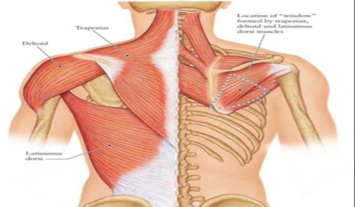
The deltoid muscle lies in the upper arms and shoulder on both sides of the body. It is majorly of triangular contour, with broad part joint to shoulder and thinnest to resting at the upper arm. It ensures flexible movement of shoulder and arms.
It is attached to three points on both sides of the body, including:
- Outer end on the collar bone (clavicle).
- Acromion process of the scapula (bony process on scapula or shoulder blade).
- The spine of the scapula (bony stature on the backside of the shoulder blade).

The Insertion And Relation of Deltoid Muscles
These are superficial fiber muscles on each side shoulder, that overlying fascia, platysma muscle, and skin.
Deltoids overlie within several other muscular structures such as rotator cuff muscles like supraspinatus, infraspinatus, teres minor, and subscapularis.
Muscles, including pectoralis major, pectoralis minor, and triceps branchii muscles. It even contours coracoacromial ligament, subacromial bursa, bony structures, and neurovascular structures on shoulders. It is innervated by the axillary nerve (brachial plexus).
Deltoid Muscles Blood Supply
The blood supply is regulated by many sources such as below:
- Thoracoacrominal Artery, a branch of the axillary artery, a combination of acromial and deltoid branches.
- Subscapular Artery, also the branch of the axillary artery.
- Anterior Circumflex Humeral Artery.
- Posterior Circumflex Humeral Artery.
- Deep Brachial Artery or profunda brachii artery from the deltoid branch.
Deltoid Muscle Function
Precisely, the deltoid muscle is the principal abductor of the arm lies on the glenohumeral joint.
It ensures easy abduction motion as made up of clavicular and scapular spinal fibers. While carrying heavy objects, deltoid muscles produce a line of force to stabilize the glenohumeral joint.
This primary prevents pain and displacement of the glenohumeral joint and controllers the arm and shoulder pressure. It even undergoes with eccentric contraction on deltoid muscles.
Clavicular-anterior fibers will help the deltoid muscle produce flexion of the arm, along with pectoralis motion while walking and running.
However, the scapular spinal fibers-posterior of deltoid muscles produce an extension of the arm while acting with latissimus dorsi during ambulation helps in the humerus’ rotation.


